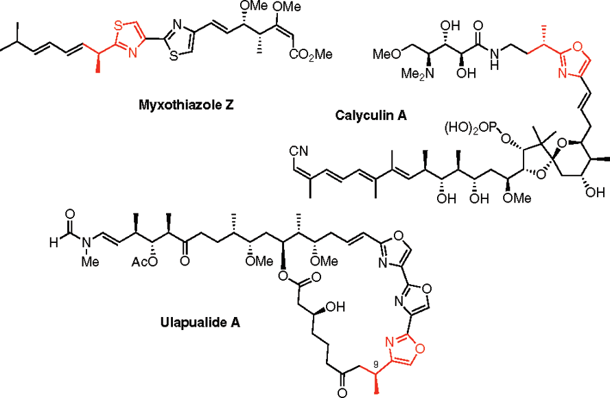
Calyculin A
CAS number: 101932-71-2
Calyculin A from Discodermia calyx is a dual action toxin that blocks calcium influx and inhibits protein Ser/Thr phosphatases.
Related images
Related Questions and Answers
Q: How does calyculin A (cal A) affect the kinetics of DNA repair foci formation in irradiated cells?
A: In a study, cal A (1 and 10 nM) did not stabilize γH2AX foci 2 hours post-irradiation. Instead, 10 nM cal A significantly decreased the number of all types of DNA repair foci (γH2AX, 53BP1, and colocalized γH2AX/53BP1) induced by high-dose (100 cGy) irradiation. This suggests that cal A may induce apoptosis-related changes in chromatin structure, preventing DNA repair proteins from accessing DSB sites.
A: Cal A (10 nM) induced apoptosis in UCBL within 2 hours of treatment, independent of the delivered radiation dose. Apoptosis was also detected in UCBL treated with 1 nM cal A at longer incubation times (20 and 44 hours). The study suggests that cal A-induced apoptosis may underlie the failure of cal A to maintain radiation-induced γH2AX foci.
A: In a study, cal A (1 and 10 nM) did not stabilize γH2AX foci 2 hours post-irradiation in UCBL. Instead, 10 nM cal A significantly decreased the number of γH2AX foci induced by high-dose (100 cGy) irradiation. This effect was attributed to cal A-induced apoptosis, which may alter chromatin structure and prevent DNA repair proteins from accessing DSB sites.
A: In Hs-68 fibroblasts, Caly A blocks serum-induced calcium influx, while in MDA-MB-468 breast cancer cells, it does not. This suggests that breast cancer cells may utilize alternative calcium entry pathways that are not affected by Caly A.
A: A study shows that treatment with up to 1 nM Caly A does not significantly affect cyclin D1 levels in either cancerous or non-cancerous cell lines. This indicates that the cell cycle arrest caused by low-dose Caly A is not due to cyclin D1 ablation.
A: Acute application of 0.3 nM Caly A blocks serum-induced increases in intracellular calcium levels in Hs-68 fibroblasts but not in MDA-MB-468 breast cancer cells. This suggests that Caly A selectively blocks calcium influx in fibroblasts.
A: Breast cancer cells are resistant to low-dose Caly A because they likely use different calcium entry pathways compared to fibroblasts. Caly A blocks calcium uptake via non-selective cation channels in fibroblasts, but breast cancer cells may rely on other channels unaffected by Caly A.
A: Long-term exposure to 0.3 nM Caly A prevents G1 to S phase progression in Hs-68 fibroblasts and ARPE-19 epithelial cells, indicating cell cycle arrest in these non-cancerous cell lines.
A: Caly A acts as both a channel blocker and a protein phosphatase inhibitor. At subnanomolar doses, it blocks calcium uptake by fibroblasts, preventing G1 to S phase cell cycle progression, while higher doses inhibit protein phosphatases.
A: Calyculin A, an established inhibitor of type 1/2A serine/threonine phosphatase activity, dramatically inhibits the Ca2+ release-activated Ca2+ (CRAC) entry pathway in intact rat basophilic leukemia (RBL) cells, a finding that re-evaluates its crucial role in controlling CRAC pathway activation and maintenance, contrary to previous reports.